INDIRECT OPTHALMOSCOPE

Rated 5.00 out of 5 based on 1 customer rating
(1 customer review)

SKU: WCM35682 Category:

INDIRECT OPTHALMOSCOPE

The Vantage Plus head-worn binocular indirect ophthalmoscope is our best-in-class offering, providing eye care professionals worldwide with high quality optics and brilliant retinal illuminations.

INDIRECT OPTHALMOSCOPE

INDIRECT OPTHALMOSCOPE
INDIRECT OPTHALMOSCOPE supplier UAE
INDIRECT OPTHALMOSCOPE supplier Abu Dhabi
INDIRECT OPTHALMOSCOPE supplier Dubai
INDIRECT OPTHALMOSCOPE supplier Sharjah
INDIRECT OPTHALMOSCOPE supplier Ajman
INDIRECT OPTHALMOSCOPE supplier Ras Al Khaimah
INDIRECT OPTHALMOSCOPE supplier Fujairah
INDIRECT OPTHALMOSCOPE Umm Al Quwain
An indirect ophthalmoscope is a specialized optical device used by ophthalmologists and optometrists to examine the interior structures of the eye, particularly the retina, optic nerve, and vitreous humor. Unlike the direct ophthalmoscope, which provides a narrow and magnified view of the central retina, the indirect ophthalmoscope offers a much wider field of view, allowing for detailed visualization of both the central and peripheral retina. This makes it particularly useful for diagnosing conditions such as diabetic retinopathy, retinal detachment, retinal tears, tumors, and other abnormalities that may not be visible through direct ophthalmoscop
The functioning of an indirect ophthalmoscope relies on a light source, a condensing lens, and a system of mirrors and prisms that allow the examiner to view an inverted image of the retina. The device is typically mounted on a headgear worn by the examiner, allowing them to have both hands free to manipulate the condensing lens and examine different parts of the retina effectively. The binocular indirect ophthalmoscope (BIO), the most commonly used type, provides a stereoscopic or three-dimensional view, which helps in assessing the depth and contours of retinal lesions, making it an invaluable tool for retinal examinations. The examiner holds a powerful convex lens, typically 20 diopters (D), 28D, or 30D, at a distance from the patient’s eye to converge and focus the light, forming an inverted and magnified image of the retina. This technique is particularly advantageous in cases where a patient has media opacities, such as cataracts, since it provides a clearer view of the retina despite the obstruction
One of the primary advantages of an indirect ophthalmoscope is its ability to examine the peripheral retina, which is crucial for detecting conditions like retinal tears, detachments, and degenerations that may not be visible with a direct ophthalmoscope. This makes it particularly useful for patients with symptoms like flashes, floaters, or sudden loss of vision, as well as those at high risk of retinal disease, such as individuals with diabetes, high myopia, or a history of trauma. Additionally, indirect ophthalmoscopy can be performed even in situations where the patient is uncooperative, such as in infants, unconscious individuals, or those with poor fixation, making it an essential tool for pediatric anemergency eye care
Despite its numerous advantages, the indirect ophthalmoscope has some limitations. The image seen through the device is inverted and laterally reversed, meaning the examiner must mentally adjust for this orientation when analyzing retinal structures. Additionally, the examination requires pupil dilation for optimal visualization, which may cause temporary light sensitivity and blurred vision in the patient. Some individuals may also find the bright light used in the procedure uncomfortable. The technique itself requires skill and experience, as the examiner must carefully align the condensing lens and light source to obtain a clear and detailed view of the retina
The indirect ophthalmoscope has revolutionized retinal examinations and disease diagnosis by providing a wide field of view, enhanced depth perception, and the ability to examine the peripheral retina, which is not possible with traditional direct ophthalmoscopy. It remains a cornerstone in ophthalmic practice, particularly in the diagnosis and management of retinal diseases, and is widely used by specialists worldwide
Eye Equipment: Essential Tools for Vision Examination and Treatment
Eye equipment refers to the various specialized instruments used by ophthalmologists, optometrists, and eye care professionals to examine, diagnose, treat, and manage conditions related to the eyes. These tools range from simple hand-held devices to complex, high-tech machines that provide detailed imaging and analysis of the eye’s internal and external structures. The field of ophthalmology relies heavily on these instruments to assess vision, detect abnormalities, and perform surgical procedures to restore or enhance eyesight
One of the most commonly used pieces of eye equipment is the ophthalmoscope, which is designed to examine the retina, optic nerve, and other internal structures of the eye. There are two main types: the direct ophthalmoscope, which provides a magnified, upright image of a small section of the retina, and the indirect ophthalmoscope, which offers a wider, inverted view of the retina, particularly useful for examining the peripheral retina. The indirect ophthalmoscope is often head-mounted and used in conjunction with a powerful convex lens, making it ideal for detecting retinal detachments, diabetic retinopathy, and other posterior powered microscope combined with an adjustable, narrow beam of light. The slit lamp allows eye care professionals to examine the anterior and posterior segments of the eye, including the cornea, lens, iris, and vitreous humor. This instrument is invaluable for diagnosing conditions such as cataracts, corneal ulcers, glaucoma, and conjunctivitis. By using additional lenses, the slit lamp can also be adapted for a more detailed view of the retina and optic nerveINDIRECT OPTHALMOSCOPE INDIRECT OPTHALMOSCOPE INDIRECT OPTHALMOSCOPE INDIRECT OPTHALMOSCOPE INDIRECT OPTHALMOSCOPE INDIRECT OPTHALMOSCOPE INDIRECT OPTHALMOSCOPE INDIRECT OPTHALMOSCOPE INDIRECT OPTHALMOSCOPE INDIRECT OPTHALMOSCOPE INDIRECT OPTHALMOSCOPE INDIRECT OPTHALMOSCOPE INDIRECT OPTHALMOSCOPE INDIRECT OPTHALMOSCOPE INDIRECT OPTHALMOSCOPE INDIRECT OPTHALMOSCOPE INDIRECT OPTHALMOSCOPE INDIRECT OPTHALMOSCOPE INDIRECT OPTHALMOSCOPE INDIRECT OPTHALMOSCOPE INDIRECT OPTHALMOSCOPE INDIRECT OPTHALMOSCOPE v v INDIRECT OPTHALMOSCOPE INDIRECT OPTHALMOSCOPE INDIRECT OPTHALMOSCOPE INDIRECT OPTHALMOSCOPE INDIRECT OPTHALMOSCOPE INDIRECT OPTHALMOSCOPE INDIRECT OPTHALMOSCOPE INDIRECT OPTHALMOSCOPE INDIRECT OPTHALMOSCOPE INDIRECT OPTHALMOSCOPE
For measuring intraocular pressure (IOP), which is a key factor in diagnosing glaucoma, tonometers are used. The Goldmann applanation tonometer, often attached to a slit lamp, is considered the gold standard for IOP measurement, providing accurate readings by gently flattening the cornea. Other types of tonometers include the non-contact (air puff) tonometer, which uses a burst of air to measure eye pressure without direct contact, and the tonopen, a handheld device that is particularly useful for children or non-cooperative patients
To assess a patient’s refractive errors and determine their prescription for glasses or contact lenses, phoropters and autorefractors are widely used. A phoropter is a mechanical device that contains multiple lenses, allowing the examiner to fine-tune a patient’s vision correction by switching between different lens powers while the patient reads an eye chart. An autorefractor, on the other hand, provides an automatic, computerized measurement of refractive error by analyzing how light is reflected by the retina. These tools help optometrists prescribe accurate corrective lenses for conditions such as myopia (nearsightedness), hyperopia (farsightedness), astigmatism, and presbyopia
For detailed imaging of the retina and other internal structures, optical coherence tomography (OCT) is a highly advanced diagnostic tool. OCT uses light waves to create cross-sectional images of the retina, allowing for the detection of retinal diseases such as macular degeneration, diabetic retinopathy, and glaucoma. The high-resolution images produced by OCT provide invaluable information about the thickness and integrity of different retinal layers, enabling early diagnosis and monitoring of progressive eye conditions
Another essential piece of eye equipment is the fundus camera, which captures high-quality images of the retina, optic nerve, and blood vessels. This is especially useful for documenting changes over time in patients with diabetic retinopathy, macular degeneration, and hypertensive retinopathy. A more advanced version of this technology is fluorescein angiography, where a fluorescent dye is injected into the bloodstream, allowing the camera to capture images of blood flow in the retina, helping to diagnose and monitor vascular abnormalitie
For corneal assessments, pachymeters and topographers are used. A pachymeter measures the thickness of the cornea, which is crucial for evaluating patients before LASIK surgery or monitoring those with keratoconus or glaucoma. Corneal topography, on the other hand, creates a detailed map of the cornea’s surface curvature, essential for fitting contact lenses and diagnosing irregular corneal conditions
In surgical settings, microsurgical instruments and laser technology play a vital role in vision correction and treatment. Excimer lasers are used in LASIK and PRK procedures to reshape the cornea and correct refractive errors, while femtosecond lasers assist in cataract surgery by making precise incisions and softening the lens for easier removal. Other surgical tools include vitrectomy machines, which are used to remove the vitreous humor in cases of severe retinal disorders, and cryotherapy probes, which are used to treat retinal tears by freezing affected areas
Pediatric eye examinations also require specialized equipment such as retinoscopes, which help determine a child’s refractive error by analyzing how light reflects off the retina. Portable vision screening devices are used in schools and pediatric clinics to quickly assess vision in young children who may not be able to communicate their vision problems clearly
Overall, eye equipment has revolutionized the way eye care professionals diagnose, monitor, and treat a wide range of ocular conditions. From simple tools like visual acuity charts to sophisticated imaging machines like OCT and fundus cameras, these instruments ensure that eye diseases are detected early and managed effectively. As technology continues to advance, newer and more precise equipment is being developed, improving the accuracy of eye examinations and enhancing treatment outcomes for millions of patients worldwideyDental equipment refers to the wide range of instruments and machines used by dentists, dental hygienists, and oral surgeons to examine, diagnose, treat, and maintain the health of a patient’s teeth, gums, and overall oral cavity. These tools are essential for performing routine check-ups, preventive care, restorative treatments, and complex surgical procedures. Modern dentistry relies heavily on advanced equipment to improve accuracy, efficiency, and patient comfort during dental procedure
One of the most fundamental pieces of dental equipment is the dental chair, which is designed to provide comfort and proper positioning for the patient while giving the dentist easy access to the oral cavity. These chairs are often adjustable and come equipped with a built-in control panel that allows the dentist to modify the chair’s height and inclination. Many modern dental chairs also include an overhead light, a spittoon, and attachment points for essential instruments such as handpieces, suction devices, and air-water syringes
A critical component of any dental examination is the dental mirror, which allows the dentist to view hard-to-reach areas of the mouth. This small, angled mirror helps in examining the back surfaces of the teeth and detecting cavities, plaque buildup, and other abnormalities. In addition to the mirror, dentists use explorers and probes, which are sharp, pointed instruments designed to detect decay, tartar deposits, and other irregularities on the tooth surface. The periodontal probe, in particular, is used to measure pocket depths around the teeth, helping diagnose gum diseas
For removing plaque, tartar, and calculus from teeth, scalers and curettes are essential tools. These instruments come in different shapes and sizes, allowing the dentist or hygienist to clean both the visible tooth surfaces and the areas beneath the gumline. In modern dental practices, ultrasonic scalers are widely used, which utilize high-frequency vibrations and water spray to effectively remove hardened deposits with minimal discomfort to the patient
Restorative procedures, such as fillings and crowns, require the use of dental handpieces, commonly known as dental drills. These high-speed and low-speed handpieces are powered by air turbines or electric motors and are used to remove decayed tooth material, shape cavities, and prepare teeth for restorations. High-speed drills are primarily used for cutting and shaping, while low-speed drills are used for polishing and finishing. Along with handpieces, burs—small, rotating cutting instruments made of carbide or diamond—are used for precise cutting and shaping of enamel and dentin
In procedures such as root canals, endodontic instruments are used to treat infections within the tooth’s pulp. Endodontic files and reamers are small, flexible metal tools used to clean and shape the root canals before filling them. These instruments help remove infected tissue and smooth the canal walls to ensure a proper seal with the root canal filling material. Advanced root canal procedures may also involve the use of dental apex locators, which help determine the precise length of the root canal to ensure complete cleaning and sealing
For tooth extractions and surgical procedures, dental forceps and elevators are indispensable. Forceps are used to grip and extract teeth, while elevators help loosen the tooth from the surrounding bone before extraction. These instruments come in various designs, each tailored for different types of teeth, such as molars, premolars, or incisors. In complex cases, such as impacted wisdom teeth, dentists may use surgical drills, bone chisels, and suturing materials to perform precise surgical extractions
Dental radiography plays a crucial role in diagnosing hidden dental problems. Dental X-ray machines allow dentists to capture detailed images of teeth, roots, and surrounding bone structures. There are different types of dental X-rays, including bitewing X-rays, which help detect cavities between teeth, periapical X-rays, which provide a full view of a single tooth, and panoramic X-rays, which capture a comprehensive image of the entire mouth. More advanced imaging techniques, such as cone beam computed tomography (CBCT), provide three-dimensional views of the oral structures, assisting in implant placement, orthodontic treatment planning, and complex surgical procedures
Dental impressions are essential for creating accurate molds of a patient’s teeth and gums. These impressions are taken using dental impression trays and materials such as alginate or silicone-based compounds. They are used in procedures such as crown and bridge fabrication, orthodontic treatments, and the creation of dentures. In modern dental practices, digital scanners are increasingly replacing traditional impression materials, allowing for faster, more precise 3D imaging of the patient’s oral cavity
For aesthetic and restorative treatments, dental lasers are becoming more popular. These lasers are used for procedures such as gum contouring, cavity removal, teeth whitening, and even minor surgical procedures. Dental lasers offer several advantages, including reduced bleeding, faster healing times, and greater precision compared to traditional surgical methodsse

Brand

KEELER

The primary goal of Keeler is to prevent vision loss by making safe, legal, and high-quality medical devices available to our customers and clients. keeler products meet all applicable regulatory requirements, including UK Medical Device Regulation 2002, Medical Device Directive 93/42/EEC, and Medical Device Regulation 2017/745. By affixing the CE and UKCA markings to our products, we certify that they are safe and meet all requirements for environmental, health, and safety. certifications include ISO 13485:2016, ISO 14001, Medical Device Single Audit Programme (MDSAP), UKCA and CE.

LUXAMED

LUXAMED ,in which we engage our suppliers from the first moment. Only high quality materials such as high-strength, polished, corrosion- and heat-resistant stainless steel or polished anodized aluminum are used in our products. However, our most important principle is still: handmade. Our product range currently comprises the following product groups and is constantly expanding. To all our products, we are making high demands on quality, high quality materials and environmental protection. products Otoscopes
Ophthalmoscopes Diagnostic Sets Dermatoscopes Penlights Stethoscopes Reflex Hammers Examination Lamps LED Examination Lamp FOCUS Focusable Tuning Forks Laryngoscopes

1 review for INDIRECT OPTHALMOSCOPE

  1. Rated 5 out of 5

    Joseph VT

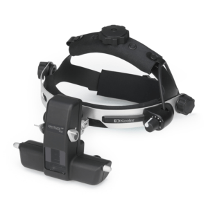
    Excellent product ⭐️⭐️⭐️⭐️⭐️
    The Vantage Plus head-worn binocular indirect ophthalmoscope is our best-in-class offering, providing eye care professionals worldwide with high quality optics and brilliant retinal illuminations.

Add a review

Your email address will not be published. Required fields are marked *

Shopping Cart